Saturday, September 14, 2013

7.5 Proteins

Four levels of protein structure
1. Primary Structure = amino acid sequences
   a) These are polypeptide chains (between 50-1000 amino acids in length)
   b) 20 amino acids
   c) R groups of amino acids aid in shaping the protein

Primary Structure
   a) The primary structure forms a –N-C-C-N-C-C-N-C-C- backbone to the molecules
   b) The primary structure is read from the NH2 – terminal to the –COOH terminal
   c) Each amino acids is identified by its specific R group
Secondary Structure
1. Secondary Structure = describes the shape of the protein
   a) There are two main types:
      i) Alpha helix
      ii) Beta pleated sheets
   b) Both stabilized by hydrogen bonding between groups in the main chains
   c) The primary structure of a polypeptide has group projecting from the N-C-C backbone
   d) There are three noted forms of secondary structure

Secondary Structure- Alpha Helix
   a) Formed from hydrogen bonds
   b) This is drawn as a helix that follows the –N-C-C-N-C- backbone of a polymer
   c) Alpha helices are often the basis of fibrous polymers (i.e. collagen)
   d) Right handed helix
   e) The alpha helix was first discovered by Linus Pauling
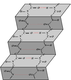
Secondary Structure- Beta-Pleated Sheet
   a) Beta-pleated sheets are so called because of the ‘pleated’ or folds when viewed from the side
   b) The polypeptide chain is much more stretched out in comparison to the alpha helix
   c) This ‘sheet’ often has twists that increase the strength and rigidity of the structure
   d) This beta-pleated sheet was discovered by Pauling and Corey


Structure- Open Loops
   a) Alpha helices and beta-pleated sheets are often connected together by short chains of amino acids which form neither of the previous structures but simply link other sections together
   b) They are in fact often important regions of proteins including the active sites of enzymes


Tertiary Structure
   a) The overall shape of conformation of a polypeptide.  Basically its just referring to the folds in a polypeptide chain
   b) These folds are formed just after translation
   c) Caused by bonding of the R groups together
   d) Hydrophilic R groups bond to each other
   e) Hydrophobic R groups bond to each other
   f) Types of intramolecular bonds may include covalent, ionic, hydrogen, disulphide bridges, and hydrophobic interactions

Tertiary Structure- Disulphide Bridges
   a) R groups that have sulfer will form covalent bonds between one another (i.e. between two adjacent cysteine amino acids).  This forms a disulphide bridge.
   b) The covalent bond stabilizes the tertiary shape of a protein


Quaternary Structure
   a) A number of tertiary polypeptides joined together
   b) Hemoglobin is a quaternary structure
   c) It is composed of 4 different polypeptide chains
   d) Each chain forms a tertiary structure called a hem (haem) group
   e) Prosthetic groups: proteins are often bond to inorganic groups (e.g. Hemoglobin has four polypeptide ‘hem’ groups each associated with Fe2+.)
Fibrous Proteins
   a) Are water insoluble, long and narrow proteins
   b) Are associated with providing strength and support to tissue
   c) Collagen is the basis of the connective tissue and is composed of three left handed helices.  They make up the extracellular matrix and are found in cartilage, ligiments, tendons, etc.
   d) This is the most common protein in animals
   e) Keratin is another common fibrous protein which is composed of seven helices (major protein in hair and nail structure)


Globular Protein
   a) Are near soluble (colloids)
   b) They have more compact and rounded shape
   c) Are associated with functions such as: pigments and transport proteins (hemoglobin, myoglobin, lipoproteins)
   d) Immune system (immunoglobins)
   e) Structural motifs- sophisticated method of describing protein structure
   f) Examples are enzymes, hormones, hemoglobin and immunoglobin (antibodies)


Significance of Polar and Non-Polar amino acids
   a) Polarity of amino acids depends on the R groups
   b) Polar amino acids have hydrophilic R groups
   c) Non-Polar amino acids have hydrophobic R groups

Polar A.A.
   a) Water soluble
   b) In the cell membrane:
      i) They create channels in proteins for hydrophobic substances to pass through
      ii) They cause part on the membrane proteins to protrude from the cell membrane
      iii) Transmembrane proteins have two polar regions (one on surface and one in channel)
   c) Cell membrane proteins:
      i) The sections of the molecule that contain polar amino acids are hydrophilic and can exist in contact with water.
      ii) Polar amino acids allow the positioning proteins on the external and internal surface of a cell membrane.  Both cytoplasm and tissue fluid are water based regions.

Non-Polar A.A.
   a) Water insoluble
   b) They stabilize the entire protein when found in the center of the water soluble amino acids
   c) They cause proteins to remain embedded in the cell membrane

Polar v. Non-Polar
   a) The lining of the channel itself will be of polar amino acids to allow the diffusion of charged molecules and ions
   b) Polar amino acids within the active site of an enzyme allow a chemical interaction between the substrate and the enzyme to form an activated complex
Four Functions of Protein:
1. Hormones (globular)-
   a) Insulin is a hormone that reduces blood sugar
   b) Produced in the beta- cells of the pancreas islets
   c) Main target tissue is muscle cells and liver cells
   d) Function: to bring about the update of glucose across the cell membrane and the storage of glucose as the insoluble polymers glycogen


2. Immunoglobulins (globular):
   a) Known as antibodies
   b) Produced by the plasma cells in an immune response to an infectious antigen
   c) Great variation exists in the heavy chains which allows a response to virtually any possible antigen surface
3. Enzymes (globular):
   a) Enzymes reduce the energy of activation and allows biochemical reaction to reach equilibrium more quickly
   b) Enzymes are large globular proteins often with prosthetic groups
   c) The maximum number of substrate molecules that can be converted into product per second (excess substrate) is called the ‘turn-over rate’


4. Gas Transport (globular):
   a) Hemoglobin molecules aid in binding oxygen to red blood cells then transporting it to respiring tissues
   b) They are contained within the erythrocytes (red cells) of the circulatory system
   c) Composed of 4 hem groups each associated with a prosthetic Fe2+ ion
   d) Each hem group can carry an oxygen atom


Proteins contd.
   a) Collagen (fibrous): provides structure for the skin
   b) Actin and Myosin (fibrous): aids muscle contraction
   c) Fibrin (fibrous): aids in the clotting of blood

Saturday, September 7, 2013

Proteins & Protein Synthesis

I.             Proteins
a.       have a variety of functions
                                                              i.      some will be used in the membrane of the cell
                                                            ii.      remain in the cytoplasm or be transported out of the cell
b.      many copies of a protein can be made from one mRNA molecule
c. enzymes, hormones are examples of proteins
II.          Protein synthesis
a.       accomplished through a process called translation
b.      composed of a chain of polypeptides
c.       after DNA is transcribed into a messenger RNA (mRNA) molecule during transcription, the mRNA must be translated to produce a protein
d.       mRNA along with transfer RNA (tRNA) and ribosomes work together to produce proteins.
III.        Transfer RNA 
a.       transfer RNA is shaped like a clover leaf with three loops
b.      contains an amino acid attachment site on one end and a special section in the middle loop called the anticodon site
c.       translate the message within the nucleotide sequence of mRNA to a specific amino acid sequence
d.      sequences are joined together to form a growing polypeptide chain
IV.           Ribosomes
a.       consist of two parts, a large subunit and a small subunit, contain a binding site for mRNA and two binding sites for tRNA located in the large ribosomal subunit
b.      clusters of ribosomes that translate a single mRNA sequence are called polyribosomes or polysomes.
V.            Ribosomes in Protein Synthesis
a.       initiator tRNA resides in one binding site of the ribosome called the P site, leaving the second binding site, the A site, open
b.      when a new tRNA molecule recognizes the next codon sequence on the mRNA, it attaches to the open A site
c.       peptide bond forms connecting the amino acid of the tRNA in the P site to the amino acid of the tRNA in the A binding site
d.      as the ribosome moves along the mRNA molecule, the tRNA in thP site is released and the tRNA in the A site is translocated to the P site
e.       A binding site becomes vacant again until another tRNA that recognizes the new mRNA codon takes the open position.

Tuesday, September 3, 2013

Transcription and Translation


7.3 Transcription

Nucleosides vs. Nucleotides
  •         A nucleoside: has a nitrogen base linked by a glycosidic bond to C1’ of a sugar (ribose or deoxyribose) (without the phosphate group)
  •         A nucleotide is a nucleoside that forms a phosphate ester with the C5’ –OH group of a sugar (ribose or deoxyribose).
  •         Phosphodiester bond join nucleotides

       Nucleoside                                                                         Nucleotide


Types of RNA
       Messenger RNA (mRNA)- carries genetic information from DNA to cytosol
       Ribosomal RNA (rRNA)- Most abundant; makes up ribosomes
       Transfer RNA (tRNA)- Binds to specific Amino Acids

RNA
       Transcription- the copying of the base sequence of a gene (DNA) by making an RNA molecule.
       Complementary base pairing rules are followed CºG except that A=U

Transcription
  •         5’ end of free nucleotides are added to the 3’ end
  •        The sense strand (coding strand) has the same base sequence as mRNA with uracil instead of thymine. – 5’ to 3’
  •        The antisense (template) strand is transcribed


Transcription Process

Initiation Explained
       The promoter region is for the binding of RNA polymerase
       This allows RNA Polymerase to:
       Find the Anti-sense strand, know the direction of transcription, and the start for transcription.
       The hydrogen bonds of the DNA helix are opened by the DNA Helicase.
       The bases of the anti-sense strand (3’-5’ for DNA) are exposed.
       RNA Nucleotides complementary base pair with the anti-sense nucleotide bases
       The free nucleotides (nucleoside triphosphates) are based on RNA.  The sugar is Ribose.
       The nucleotides are Adenine, Guanine, Cytosine and Uracil.    

Elongation Explained
  •         The RNA polymerase forms covalent bonds between nucleotides (phosphodiester).
  •         Free energy is released from the oxidation reaction of the nucleoside triphosphates to form the bond.
  •      The bonds are formed by joining the 5’ of the free nucleotide to the 3’ end of the nucleotide already part of the mRNA chain
  •      The RNA polymerase works along the nucleotides completing the ribose-phosphate backbone


Steps of Transcription
  1. Helicase uncoils the DNA by breaking the hydrogen bonds of the complementary base pairs at the position of the gene.
  2. RNA polymerase finds the promoter region on the antisense strand of DNA (TAC)- Initiation.
  3. Free RNA nucleotides complementary base pair with DNA nucleotides on the antisense strand. A=U, GºC
  4. The phosphodiester bonds on the mRNA chain are formed by RNA Polymerase- Elongation.
  5. The RNA polymerase reaches the terminator (DNA- ATT, ATC, or ACT) and the RNA polymerase stops.
  6. The mRNA is complete the molecule detaches from the DNA and leaves the nucleus for the cytoplasm ribosomes.
  7. The DNA helix reforms

SNRPS
       Are used to remove introns to form mature mRNA
       Pre-mRNA has been produced through transcription of the anti-sense strand as described for prokaryotic transcription.
       (a) The non-coding introns are spliced out of the mRNA.
       The introns are broken down in the nucleus.
       (b) The remaining mRNA is called mature mRNA and is exported from the nucleus to the cytoplasm for translation into the polypeptide.

7.4 Translation

One Gene, One Polypeptide
       Theory- One gene is transcribed and translated to produce one polypeptide.
       Some proteins are composed of a number of polypeptides and in this theory each polypeptide has its own gene.
       e.g. Hemoglobin is composed of 4 polypeptides (2 of each type) and there is a gene for each type of. polypeptide.

Codons
       Genetic code- A T(U) G C, is used by most organisms to translate mRNA into proteins.
       Codon- 3 (triplet) nucleotides of mRNA that code for an amino acid.
       Anticodon- 3 nucleotides of tRNA that are complementary to and pairs with the mRNA codon. They carry the amino acid.
       A polypeptide is a sequence of bases
       Bases are either A,T,G, or C
       64 codons code for 20 amino acids.
       Less of a chance for mutations
       The genetic code is universal so it is the same in almost all organisms
       Each codes for the addition of an amino acid to a growing polypeptide chain
       The genetic code is degenerate- meaning more than one codon can code for a particular amino acid
       AUG is the start codon
       Some codons code for the end of translation- Terminator codons (UGA, UAA, UAG)


Translation
       The location of translation is the ribosomes in the cytoplasm.
       Ribosomes are composed of rRNA which acts as a catalyst for the translation of mRNA.
       mRNA from the nucleus locates a ribosome.
       The start codon (AUG) occupies one of two ribosome sites.
       The ribosome moves along the mRNA
       One mRNA can have many ribosomes (polysome) which accelerate protein synthesis.

tRNA
  •  Each amino acid has a specific tRNA-activating enzyme (aminoacyl-tRNA synthetase)

       tRNA is composed of one chain of (RNA) nucleotides
       tRNA has an anticodon
       Anticodon of three bases which are single stranded and form part of a loop
       tRNA has double stranded sections formed by base pairing
       tRNA has 3 loops (sometimes with an extra small loop)
       tRNA has a distinctive 3D clover leaf shape
       Activation specificity: how does the tRNA attach to the correct amino acid.
       Shape of each tRNA is different.
       Shape of the tRNA is defined by the loop and the helical sections.
       The enzyme adds a specific amino acid to the CCA base sequence (at 3' end of the tRNA) this requires ATP (energy).
       Each amino acid has one or more tRNA molecules- example of a degenerate code.


Binding Sites for tRNA
       A- Amino Acid- is the position that the new tRNA codon-anticodon binds making sure that the correct amino acid is in position.
       P- Polypeptide- is the position in which the amino acid on the tRNA adds to the polypeptide.
       E- Exit- is the position the tRNA (w/o amino acid) locates and is then released from the ribosome to become reactivated.

Translation Explanations
       Initiation: In which the ribosome, tRNA and mRNA come together to begin the translation of the mRNA.
       Elongation: tRNA molecules attach to the mRNA based on the codon-anticodon recognition. Amino acids are brought together and polymerized into the primary structure of the polypeptide.
       Translocation: The movement of the ribosome along the mRNA strand one codon at a time.
       Termination: mRNA and the ribosomes detach from one another. The polypeptide is released and the tRNA return to be charged with more amino acid.

Steps of Translation
  1. Initiation- ribosomes bind to mRNA.  Initiator tRNA binds the start codon to the small subunit of ribosome.
  2. The start codon (AUG- methionine) occupies the P site.
  3. Elongation- ribosomes moves along mRNA facilitating addition of amino acids.
  4. Second tRNA binds to ribosome at the A site.
  5. Large subunit moves down mRNA after a second tRNA binds.
  6. The amino acid on first tRNA is bonded to amino acid on second tRNA.
  7. Peptide bonds between amino acids are formed with the aid of peptidyl transferase.  Requires GTP- Guanosine-5'-triphosphate.
  8. The ribosome translocates- moves down the mRNA.
  9. The first tRNA moves to the E site and is removed and new tRNA binds.
  10. Each tRNA moves from the A site to the P site to the E site.
  11. Termination- the ribosome reaches a stop codon The stop codon causes the polypeptide to be released. 
Ribosomes
  •        free ribosomes synthesize proteins for use primarily within the cell
  •        bound ribosomes synthesize proteins primarily for secretion by lysosomes
  •     Proteins and Ribosomal RNA combine in the structure.
  •        Large sub-unit has three binding sites for tRNA molecules (E, P and A site).
  •        Small sub-unit has a binding site for mRNA.
  •        Ribosome Function: Ribosomes are enzymes that catalyze the translation of mRNA into a polypeptide- their substrate is mRNA


Peptide Bond
                           

Comparing Transcription and Translation